Tuesday, May 31, 2016

Cray fish dissection

Crayfish are freshwater crustaceans that live in streams, rivers, and lakes. While they can be found practically everywhere in the world with these conditions, crayfish are most abundant in the United States/ North America. Crayfish eat decaying plants, dead fish or insects, and living plants. Like fish, crayfish have gills that they breathe through in order to obtain oxygen that is then dissolved into their bloodstream. One fun fact is that there are many different unique colors of crayfish that can be found such as bright blue, green, and orange.


Internal:
Green Gland: Digestive enzyme that produced digestive glands
Flexor Muscles: Help move the body of the crayfish and maintain coordination in water
Anterior Gastric Muscle: Help the stomach in digestion

External:
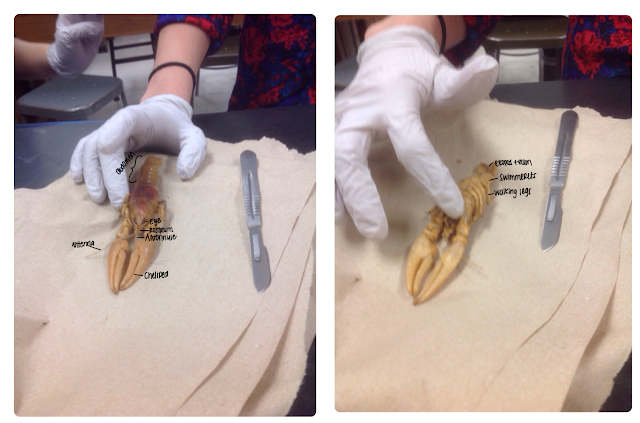
Cheliped: Used for defense and to capture prey
Walking legs: help the crayfish move from one location to another
Swimmerets: Abdominal appendage that help move water
Antennae: Senses taste and touch
Incision guide


Video link

Clam dissection

Clams are a part of the Mollusca phylum and the Animalia kingdom. Different species can be found in both salt and freshwater environments. They can often be found buried in the sand or in the mud. Clams eat plankton in order to gain energy and survive. They are able to eat them by pumping water containing the plankton through their gills. Clams breathe through their gills just like fish do. One fun fact about clams is that they do not have eyes, ears, or a nose!


Internal:
Foot: Used to dig down into the sand and draw nourishment.
Gonad: Produce gametes
Gills: Help to exchange gases and provide air.
Posterior Adductor: Pulls foot back in shell

External:
Umbo: Oldest part of clam shell, it’s a bump on the shell
Mantle: Tissue that lines valves
Growth Ring: Can help determine age of clam

Incision guide

Video link 

Earthworm dissection

Earthworms are a part of the Annelida phylum, and the Lumbricidae family. Earthworms live in the soil, and because soil is so abundant around the world, they can be found pretty much anywhere. Earthworms get their nutrition from decaying things, animal feces, or decomposing animals in the soil. Worms do not breathe through lungs, and instead it absorbs oxygen from its skin. Then, oxygen is dissolved into the bloodstream so that the worm can function and survive.
One fun fact about them is that earthworms are both female and male because one earthworm produces sperm and eggs.


Internal:
1.Crop: Food is stored here for the earthworm
2. Gizzard: Food is crushed here after moving from the crop using stones.
3.Pseudohearts: Keep the blood flowing throughout the body.
4. Esophagus: Connection to crop helps move food for digestion
External:
1. Anus: Where the digested waste comes out from
2. Mouth: Entrance for food
3. Setae: Help anchor worm when moving in soil
Incision guide


Video link


https://m.youtube.com/watch?v=jEPddCPuHXQ


Perch dissection


        Perch is a freshwater fish that can be found in the lakes and rivers of North America. Their diet consists of a variety of organisms such as snails, crayfish, mussels, leeches, insects, worms, and even fish eggs. Perch breathe by taking water in through their mouths, and then passing it through its gills. When this occurs, oxygen is able to dissolve into the bloodstream. Something interesting about perch is that they are not very good swimmers because they are not able to speed up quickly and easily. 


IMG_2366.PNG


External: 
Eye: Helps the perch to see in the water
Pectoral Fins: Allows fish to tune its movement and keeps it from moving to the side. 
Nares: Nostrils help remove moisture from exhalation
Spinal Dorsal Fin: Stabilize during sudden turns
Internal:
Kidney: Helps regulate water and salt balance within the fish
Gills: Exchange gases and transfer ions.
Spleen: Reservoir for blood within the perch
Stomach: Helps with mechanical and chemical digestion.

Incision guide


Video link
https://m.youtube.com/watch?v=FKG7F6s4TCk


Frog dissection

Frog

Frogs are a part of the Amphibia family, so they live on land but reproduce in the water. They can be found on all of the continents besides Antarctica, because the living conditions are too harsh for them to survive. There are 90 species of frogs in the US today. Frogs eat insects such as moths, flies, dragonflies, and mosquitoes. It's food is swallowed through the esophagus and then is passed through the digestive tract and the digestive glands. From there it goes through the stomach and is passed through the small intestine where it can digest its food. Frogs breathe through their nostrils and larynx, which are a part of the respiratory system. They have 2 lungs which have capillaries that allow materials to pass in and out of the body. One fun fact about frogs is that they are able to jump further than 20 times the length of their body.
IMG_2370.PNGIMG_2368.PNG


Internal:
1.Lungs: Lungs help exchange gasses and filter the air to allow the frog to breathe on land.
2.Stomach: Helps to digest food (mechanical and chemical digestion).
3.Intestines: Final digestion and absorption of nutrients
4.Gallbladder: Produces bile
5. Vomer teeth: Help the frog chew
External:
1.Eyes: help the frog to see above the water and in the water
2. Nictitating membrane: helps to moisten the eye while allowing the frog to see.
3.Tympanum: Transmits sound into inner ear
Incision guide


Link to dissection video 


Friday, May 13, 2016

Artificial Selection Lab

Artificial Selection Lab
The purpose of the lab was to artificially select the traits we wanted to express in the second generation of the fast plants seeds. We wanted to see if we could pick and choose which plants it breed together to come up with a specific phenotype.


    Evolution occurs when characteristics of a population evolve over a long period of time. In natural selection, genes of a population that are favored will be passed to offspring, continuing the presence of the trait. Traits that are not beneficial to the organism, and possibly even harm them, are not passed on to the next generation and die out. This could be due to change in the environment, natural selection of genes, or another change in traits. Artificial selection is the opposite of natural selection, because the environment is not determining the traits. It is the ability of humans to alter the way in which evolution works by controlling which individuals will reproduce., therefore controlling the results of the cross. This process does not occur naturally in the environment, and can only be done by human control. By doing this, we can attempt to control or predict the traits of their offspring. Plants can be artificially selected by choosing which breeds of flowers will reproduce with another. Another example of artificial selection are the different breeds of dogs that we have. Humans have picked and altered the traits of wolves, choosing which traits offspring should have by selecting which specimen will reproduce. In this lab, we attempted to artificially select which traits the offspring of the parental generation would have. By choosing which traits we want for the offspring, we determine which parent generations to cross.


Procedures: We got a cup of dirt to put in our pot and labeled which type of plant we had to keep track of growth and so we could pick which plant to breed. We cut a wicking cord to feed it through the bottom so it could distribute water to the plant.


Next, we planted the F1 generation of seeds and monitored growth


IMG_1220.JPG


Finally, we monitored growth over a long period of time. For a while we were basically just watering dirt, but eventually our plants began to grow a little bit.


IMG_1223.JPGIMG_1347.JPGIMG_1411.JPG


Next, we went on a three day weekend and came back to all dead plants that looked something like this due to a lack of water. Possibly due to the failed wicking cord.


Lastly, after using the cheat seeds we began to see actual flowers. I think that due to us being on top of our watering after having dead plants in the reason that we saw so much growth.
IMG_5475.JPGIMG_5476.JPG


We planted F1 seeds. We planted three purple stem hairy seeds, three non purple stem yellow green leaf, and three F1 non purple hairless( this one is short). They began to grow the first couple days and weeks. After about two weeks or so they died. This could be due to lack of water. On Friday they were alive after a three day weekend they were no longer living, fear not they provided nutrients to the soil. We used the cheat seeds for the F2 generation. We did not actually artificial select the traits we wanted to see expressed in the F2 generation because all of our F1 plants died. From the F2 seeds we should have seen 9 purple stemmed green leafed, 3 purple stemmed yellow leaves, 3 green stemmed green leaves, 1 green stemmed yellow leaves. We saw one purple stemmed yellow leaved flower, one green stemmed green leaved flower, and one green stemmed yellow leaved flower. The difference in observed and expected phenotype counts could be due to some of the plants not sprouting. The plants that did sprout could have blocked other plants from sprouting. The seeds on the top of the soil could have not sprouted because they could have been displaced after we watered the plants. It could also be due to potentially defective seeds that may be old or that didn’t sprout properly because they were under light. The seeds that were too deep in the soil didn't sprout because they didn't get enough sunlight. The wicking cord could have unevenly supplied water for some plants. Human error of neglect also played a role especially in the F1 generation. The lack of water for a few days caused the F1 generation to die.  


In conclusion, this lab was not completely successful. Due to human error the plants in the F1 generation died and thus we were not able to breed the plants to come up with our own expressed traits. If done correctly, we should have seen some tall plants some plants with purple stems and some short plants and some with green stems. These would have resulted in a 9:3:3:1 ratio but some of the plants didn't grow at all. This could also be a result of defective seeds, causing the plant to not grade at all. Essentially, we should have seen a variety of phenotypes fromt the plants which we did not due human error, possibly defective seeds, or lack of water.


Saturday, April 2, 2016

PSA- Addison

PSA:
https://youtu.be/NDhN_Y1m6A0

PSA Video


(Sorry, a YouTube video was the only way I could figure out how to upload this.) 

Sunday, March 27, 2016

Common ancestry- Addison

     Studying the genetic code of organisms give biologists an idea of common ancestors among organisms. Mammals all have very similar genetic codes and only have a little bit of variance in amino acid sequence that changes their species. This may hint that we all derived from the same species. For example, scientists are able to compare the different bone structures that are similar in different species, and see where other species may have diverged from. Humans have a tailbone, but we do not have a tail. This suggests that we share ancestry with organisms who have a tail, and may have derived from a species that had one. This is because our genetic code is so similar to other mammals who have a tail.
    Membrane bound organelles are also a hint at common ancestors. There is evidence to suggest that mitochondria once was actually a prokaryotic bacterial cell. And over time it worked with and engulfed another to have one organism inside of another. This shows that the common ancestor of mitochondria and other membrane bound organelles are prokaryotes. Over time these different organisms worked together called endosymbiotic theory, and created the membrane organelles that now work in our body.
   All bacterial cells have circular chromosomes, while eukaryotic cells have linear chromosomes. This, again suggests a common ancestry among all prokaryotic organisms and al eukaryotic organisms. Because all prokaryotes have circular chromosomes, we can assume that one prokaryotes had this and passed it on for many generations. The same can be said for eukaryotes. All eukaryotic cells have linear chromosomes, and not circular. This is another reason why eukaryotic organisms are more similar gat prokaryotic ones. Prokaryotes cannot have the linear chromosomes because they did not derive from eukaryotes.
    

Sunday, March 20, 2016

Endergonic Vs. Exergonic -Ami

Endergonic: Photosynthesis is an example of an endergonic reaction. An endergonic reaction requires a high amount of activation energy to begin the process. This process would absorb free energy to store within the plants and is nonspontaneous. Nonspontaneous means that they require energy to be input in order for the reaction to occur. The overall change in free energy is positive, and this is an anabolic reaction because it builds up energy.


Exergonic: Cellular Respiration is an example of an exergonic reaction. An exergonic reaction requires a smaller amount of activation energy to start the reaction because the reaction is spontaneous. This means that they can occur without energy input. The overall change in free energy is negative, and this is a catabolic reaction reaction because it gives off energy.

These reactions interact with each other because through energy coupling the use of an exergonic process such as cellular respiration can be used to drive photosynthesis. Both of these are significant reactions because they are necessary to our survival and for plants to survive. 

Artificial Selection -Ami

Artificial selection is basically selective breeding. Humans have impacted this because we choose which traits we want to have and breed out bad traits. For example, with dogs, we have tried to breed out aggression and anger. We want dogs to be playful and happy and in order to do this we have chosen a species to breed again and again to make something more favorable.

Because dogs are kept in home their environment is around children. Because of this people have strayed from wanting aggressive dogs, they may have once used them to guard their land or protect their property but are rarely used for that purpose anymore.

In the future we may need guard dogs or we may need some animals that are aggressive but we have attempted to end these traits. Dogs are seen as house pets and in the future I believe they still will be just house pets. They are playful because of the way we have bred them over time.

Thursday, March 17, 2016

Loss of genetic diversity within a crop species- Addison

     Evolution is the change over time. As the environment changes, animals must adjust in order to survive. For example, peppered moths mushy adapt based on the trees that there are in the forest. In a light forest, lighter moths survive because they can be more easily blended in. The same is true for a dark forest but with dark moths. If the moths migrate to a forest with the opposite of their color, they will need to adjust to thier new environment and slowly edit their traits over a span of time.
      Humans are altering the environment by removing forests to build buildings, and they also contribute to global warming. This impacts the environments that different animals live in, causing them to need to change for their new environment. If they cannot adapt over time, their species will die out and become extinct
   

Monday, March 14, 2016

Restriction Mapping of Plasmid DNA

The purpose of this lab was to use restriction enzymes in order to compare the DNA using gel electrophoresis.

Introduction: Restriction mapping of DNA allows sequences of DNA to be recognizable. Restriction enzymes cut the DNA sequence as well as allow you to find the position of the DNA. You can see how far the DNA travels using the method of gel electrophoresis.  How far the segments travel on the gel relates to how long the DNA fragment is. Smaller fragments of DNA move further than longer segments. Smaller segments have less DNA, and less to “carry” so they travel further. DNA has a negative charge and is placed on the negative side of the gel. A current running through allows the DNA to travel to the positive side of the gel. Smaller bands are on the positive side of the gel, while larger pieces stay by the negative side of the gel. You can tell which fragments are smaller, larger, and which may have been been broken off from their original fragment.  
First, we got a pre made gel that had already been set and placed it into a plastic holder. This allowed us to determine which was the positive and negative end.
Next, we loaded the gel. In each well of the gel we placed the different DNA. We used 1 pipet per tube of DNA in order to extract the DNA and place it into the appropriate well without contamination. We made sure to place all of the DNA inside of the well so that it took up the entire space.

Then, the electrophoresis chamber was closed and power was run through it. After the current was applied for some time, the gel started to move from the wells to the other end of the gel. After left for some time, we removed the gels and were able to see the bands that had formed due to the current running through the gel.

    After we removed the gel from the electrophoresis chamber, we used a light box in order to examine where the DNA bands had formed.

Using a bag we marked where each of the bands had been and where the wells were. This allowed us to have a permanent source of data incase something were to happen to our gel or bands so that we could not see where they ended up.
We were then able to measure the bands and find out what the distances were from one band from the next.    

IMG_1299.PNG


Discussion: We were able to use gel electrophoresis in order to map a plasmid. There are two PstI restriction enzyme cut sites since the channel in he fell shows two markings. There are two SspI sites since the channel cut SspI creates two new fragments from one large fragment cut by PstI. There are two HpaI restriction enzyme cut sites. Since the HpaI creates new two fragments from one large fragment cut by PstI. When the DNA was cut with HpaI,HpaI/SspI, and HpaI/PstI the fragment length of 1093 was made. This means that HpaI cuts the DNA at this point and that it will remain unchanged when oh her restoration enzymes are added to HpaI. The 4700 segment cut by HpaI is cut even more when you add SspI or PstI. When you add SspI the segment is cut into pieces 1986 and 1700 long . When you add PstI the 4700 segment is cut into pieces 2140 and 514 long. Based on these observations we were able to label our plasmid to see which restriction enzymes cut where. Based on our plasmid pictured above, we decided that HPAI cuts both at 514 and 805 are further cut by SSPI which cut at 1159 and PSTI at 1900.We compared this data to our gel to see how many bands each had and compared them to the row before to determine where the next restriction enzyme would cut. We know the largest cut is PSTI because of the distance that it travels and because the other restriction enzymes do not cut that piece of DNA, it is only cut into about half which is still a large piece.

Conclusion: From restriction mapping, we are able to determine where certain restriction enzymes cut. This helps us determine the size of the fragments of DNA. From gel electrophoresis, we are able to compare a test piece of DNA to other fragments of DNA being tested. Also, on the gel by the distance that the DNA fragment travels we are able to tell the relative size. Based on the distance traveled on the gel and the other restriction enzymes we are able to draw a plasmid (pictured above) and determine the cuts in the plasmid for the relative sizes of DNA.

Friday, February 26, 2016

E. Coli Lab

The purpose of the experiment was to genetically transform E.coli to make it antibiotic resistant and glow in the dark through genetic transformation. We were also trying to make specific E. Coli glow under a blacklight.

Genetic transformations are when a gene is inserted into an organism to change that organism’s traits. Single celled organisms are better suited for transformation than multicellular organisms because of how quickly they are able to reproduce. Because bacteria is a single cell, it is able to reproduce quicker. Organisms that reproduce quickly are also better suited for transformations because you can see the results of the offspring's traits quicker. E. coli is well suited to genetically transform. E. coli becomes antibiotic resistant when you add the plasmid pGLO to it. The pGLO plasmids encodes the gene for gfp. Gfp is the gene that is resist to the antibiotic ampicillin. PGLO also has a gene regulation system that controls the expression of the fluorescent protein. The gene is switched in when it is the presence of the sugar arabinose. Transformed cells will appear white in normal lighting and fluorescent green in uv lighting.


Method
  • First we labeled one test tube -pGLO and another one +pGLO
  • Using a sterile pipet we put 250 ul of CaCl2 into each tube and put the tubes on ice
  • Then using a sterile loop we picked up an E. coli colony and put the loop into the +pGLO tube and spun the loop until all the E. coli was off of it. We repeated this with the -pGLO.
  • We then used a sterile loop to transfer pGLO plasmid from the stock tube into the +pGLO tube.
  • We out the two tubes on ice for ten minutes, and then into a hot water bath for fifty seconds and then immediately back into the ice for two minutes.
  • Using a sterile pipet we transferred 250 ul of nutrient broth to big test tubes.
  • Using a sterile pipet we transferred 100 ul of -pGLO solution onto a transformation plate with lb, and transferred another 100 ul of -pGLO solution onto a plate with lb and ampicillin
  • Using a sterile pipet we transferred 100 ul of +pGLO solution onto a transformation plate with lb and ampicillin, and transfer 100 ul of +pGLO solution onto a transformation plate with lb, ampicillin, and arabinose.
  • Using a sterile loop for each plate spread around the E. coli on the plate.         


IMG_4529.PNG

You would expect to find the most growth on the plate labeled lb,amp,ara since the E. coli is antibiotic resistance in that plate and it should have an affinity for the sugar arabinose. The amp and ara are what allows the E. coli to grow as well as glow in the dark. Any genetically transformed E. coli  would be the two plates with the +pGLO since the pGLO was added. This should cause the E. coli to be be antibiotic resistant. The plate containing the sugar should have E. coli that glows in the dark. This was the only plate that had that added. We compared the genetically transformed plates to the two plates without pGLO. E. coli should grow on the plate with no pGLO and no ampicillin to show that the E. Coli can grow on their own. There should be no growth on the plate with no pGLO and ampicillin since the ampicillin should kill the E. coli if it's not antibiotic resistant. In the problem above you can see we calculated transmittance percentage. The percentage shows how well the cells were able to express the gene. As shown, just over one half of the cells expressed the genes which is why the E. Coli was not glowing as much because we had such few cells.
In our results, we were able to see a very small amount of E. Coli glowing under the blacklight. This could be due to a lack of sugar within the dish that we placed the E. coli in. However, we were able to see a very small amount  We had successful growth in our dishes, so there was most likely no contamination of pipettes that were not sterile. If this were to happen, there would have been growth in E coli that there should not have been. The +pGLO would have mixed with the -pGLO, which could have changed our results. Since this did not occur, we saw growth and had antibiotic resistance in the correct dishes.

In conclusion, we were able to use pGLO on E. Coli in order to make it antibiotic resistant using genetic transformation.The plasmid was able to allow us to move the DNA of the E. Coli interchangeably with the gene that codes for the Green Fluorescent Protein. E  Coli was the best organism to genetically transform because it is a single celled bacteria. This allowed it to reproduce more quickly than a multicellular organism, therefore we were able to tell which coli had the most growth in the dishes. We were able to also see the efficiency at which the E. Coli expressed the genes by the transformation efficiency.

Friday, January 8, 2016

Mitosis & Meiosis Lab

3A:
Purpose: The purpose of this lab was to observe the different stages of mitosis and approximate how long the cell spends in each stage.

Introduction: Mitosis is a process that enables the replication of somatic, or body, cells. When mitosis occurs, 2 identical cells are produced.Each of these cells have 46 chromosomes. In order for mitosis to occur, it must go through a series of steps: prophase, metaphase, anaphase, and telophase. Interphase is a step before all of these and it contains a g1 step, where the cell spends most of its life growing, the S phase where replication of DNA occurs and the g2 phase where the cell prepares for prophase. Interphase also contains a G0 phase where a cell makes the decision to not undergo mitosis. During prophase, the chromatin within the cells begin to form chromosomes. In metaphase, the chromosomes formed line up in a single file line down the center of the cell. Anaphase is when the chromosomes split at the centromere, and walk along the spindle fibers to opposite sides of the cell. Finally, during telophase the cell pinches off to create two, new, identical cells.

Method: In order to see the amount of time that the cell spends in each phase, we looked at an onion root tip through a microscope.
Under the microscope we were able to clearly see the individual cells separated by the cell plate. By looking carefully at each cell, we had to determine which stage it was in by looking at where the chromosomes were located.


Discussion
From the graph above, we can see that the cell spent the longest time in interphase. The cell spent seventy three percent of the time in interphase. This is because all cells start off in interphase and some may continue on through the first checkpoint in the cell cycle and decide to divide. The cell also has to grow and get ready to did divide in interphase Other cells may not go on past the first checkpoint. The least amount of time a cell spent was in  anaphase and telophase. This may be due to the last checkpoint which occurs in metaphase. If the cell does not pass this checkpoint it will not go onto to anaphase and telophase. Usually the cell spends around ninety percent of the time in interphase. We may have had some error in the not counting all of the cells or mistaking a cell in interphase as one in prophase. The cell spent about 11% of its time in prophase, in which parent cell chromosomes condense and compact. Our data is not entirely accurate since a cell spends majority of its time in interphase. This error could have been because we didn’t count all the cells or because we mistakenly placed some in a different category. From the graph above, we can also see that telophase takes the least amount of time because it is the last phase of mitosis. During telophase, the chromosomes simply assemble at opposite ends of the cell and cytokinesis, or division of the cytoplasm, usually occurs and two identical cells are created.

Conclusion:
In conclusion, from this portion of the lab we were able to see that a cell spends the majority of its life cycle in interphase. Interphase results in the growth of cells and DNA replication in order for the cell to continue through the other steps of mitosis. A cell has to grow and have all the things it needs: organelles, DNA, correct number of chromosomes, etc. in order to go through the rest of the stages. It makes sense that each stage decreases in time because most of the growth and formation occurs in the beginning and the remaining steps do not need nearly as long because only a few things have to form and attach correctly.


3B.1
Purpose: The purpose of this lab was to show the stages of meiosis using chromosome models. These models represented a chromosome going through both Meiosis I and Meiosis II. It also is used to represent how the crossing over of genes occurs.

Introduction:
Meiosis is different from mitosis in that it produces 4 unique haploid cells rather than 2 identical diploid cells. During meiosis I, the chromosome number changes from diploid (2n) to haploid (n). In Meiosis II, the sister chromatids are separated which creates 4 haploid cells as the final product. The homologous chromosomes needed to complete this process are brought together through synapsis. After this crossing over can occur, where the different chromosomes are able to  exchange their genetic material. Calculating the the distance between two genes is based on how much crossing over occurs.

Method: For this section of the lab we used a model with beads to represent a chromosome. We were able to walk through each step of both meiosis I and II with the beads.We were also able to simulate the action of crossing over with these beads.  

Discussion:
In G1 of interphase, the chromatin is coiled within the nuclear envelope. The centrosomes are located within the plasma membrane and are close together without having formed spindles.

In prophase we begin to see the chromosomes we see the early mitotic spindle within the plasma membrane. There are still fragments of a nuclear envelope that has began to dissolve away. The sister chromatids begin to cross over and the mitotic spindle begins to form. The homologous genes cross over and the site of this is the chiasmata.


In metaphase the chromosomes begin to line up horizontally against the imaginary metaphase plate. The chromosomes are attached to the kinetochores and the mitotic spindle becomes more prominent and the centrosomes are at the top and the bottom of the cell.

In anaphase the homologous chromosomes separate and one pulls to the top while the other pulls to the bottom. The sister chromatids remain attached leading to a haploid.
In telophase the cell begins to spit in half causing a cleavage furrow. The mitotic spindles dissipate and the nuclear envelope fragments come back since there are going to be two cells and they need a new envelope.

In prophase II there are two cells with a nuclear envelope and the mitotic spindles form again in the cells. There is no DNA replication from telophase I to prophase II.
In metaphase II, the chromosomes line up vertically against the imaginary metaphase plate. The mitotic spindles form horizontally connecting to the centrosomes. The nuclear envelope dissolves.
In Anaphase II, the sister chromatids separate.

In telophase II, the two individual cells begin to split forming daughter haploid cells. The nuclear envelope forms again to protect the four new daughter cells and the mitotic spindles disappear.

Conclusion:
In conclusion, from this lab we are now able to visualize the stages of meiosis and pick out the differences between meiosis and mitosis. We also know that no chromosome replication occurs between the end of meiosis I and the beginning of meiosis II because the chromosomes are already replicated. Meiosis results in four haploid cells because the DNA splits and only half of it is taken to form cells that have combination of genes inherited from 2 parents. We are able to see that there is a strong amount of variation between people because independent assortment and crossing over which results in unique combinations of genes.

3.15
Purpose: The purpose of this portion of the lab is to examine how much crossing over occurs during meiosis.
Introduction: Crossing over is when non-sister chromatids trade parts of their DNA segments. This can happen multiple times while meiosis occurs. Because crossing over is possible, there is a greater chance for variance among gametes The crossing over of genes happens during the first part of meiosis, Meiosis I.

Method: Using the given slides, we counted and kept track of which chromosomes crossed over and which did not. When there were alternating colors, we knew that crossing over had occurred. When there were  of the same color in a row, we knew that no crossing over had occurred.
Discussion: Crossing over is dependent on the amount of distance between the genes. In order to determine the relative distance we need to use a map unit, which is an arbitrary unit that describes the distance between the linked genes. Based on the information gathered, we can understand that linked genes that are further apart have a high chance of crossing over compared to those genes that are closer together on a chromosome. We counted the ones that had four in a row which indicated no crossing over as the table indicated. The ones that showed crossing over we counted as 106. When we took the total number of Asci, which was the ones that crossed over and the ones that didn’t the total was 195. We took 106, the number that showed crossing over, and divided it by 195, the total number, and cut that number in half. We did this to find the percent showing cross over divided by two in order to find map units. The map units provided us with a distance. We drew this above to indicate the percentage of crossing over within a map unit.

Conclusion:
Genes that are further apart have a higher chance of crossing over. This is because the map unit percentage goes up and the gene is able to cross over properly because the amount of distance has increased. This leads to genetic variation because there are many different possible traits when things cross over.